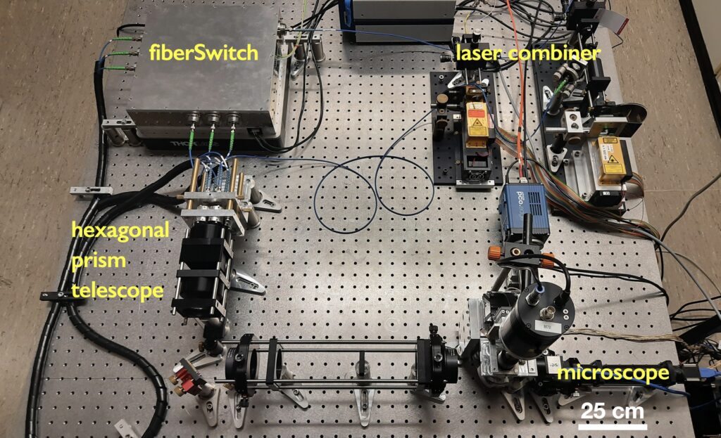
Our core imaging technology is super-resolution structured illumination microscopy (SR-SIM). In SR-SIM, mutually coherent laser beams are superposed to create an interference pattern. By implementing SR-SIM with fiber optics, we have developed an extremely flexible means to create arbitrary interference pattern and can illuminate the largest possible fields of view, as can be seen below.
The FiberSIM
Our fiber-optic implementation of SR-SIM, the FiberSIM, is a highly modular super-resolution microscope. Its main components are a laser combiner, providing rapid access to long coherence length laser sources, the fiberSwitch as the core unit providing most of the functionality of the system, the hexagonal prism telescope, which allows us to rapidly switch between imaging modalities and a very compact microscope that allows us to precisely position samples within the field of view of an objective lens with high numerical aperture (typically 60x, 1.5 NA).
FiberSwitch in action
The fiberSwitch splits laser light from the laser combiner into 3 pairs of output fibers. It is similar to a Michelson interferometer with galvanometric mirrors sending the two interferometer arms to 3 different polarization-maintaining single mode fibers, respectively.
Each interferometer arm also contains a broadband phase shifter that allows us to vary the phase of the resulting interference pattern.
The hexagonal prism telescope
The hexagonal prism telescope allows us to seamlessly change the separation between a pair of laser beams. This enables us to utilize a wide range of different SR-SIM imaging modalities – from 2D-SIM and 3D-SIM to grazing incidence SIM and TIRF-SIM.
Prism telescope in action
Collectively, by utilizing the combined functions of all the different units, we can image samples such as liver sinusoidal endothelial cells over very large fields of view (up to 150 µm x 150 µm with the 60x objective lens, and up to 250 µm x 250 µm with a 40x, 1.4 NA objective lens) at a spatial resolution of down to 95 nm very rapidly and in multiple color channels.
More details about our core imaging technology can be found in our Publications section, or in:
Multiparametric Microphysiological System (MPS) Technology
As part of work package 1 of the DeLIVERy project, Cherry Biotech are working to develop the first multiparametric Microphysiological System (MPS) platform for the culture and treatment of patient-specific cells for use with the biological models developed within the project.
There are 3 main objectives of this platform: 1. A user-friendly, long-term environmental control platform for complex MPS models (such as liver biopsies, spheroids or vascularized liver endothelial 3D ensembles) 2. To tightly control the biophysical parameters (shear stress, dissolved o2, pH, temperature) 3. Integration with imaging modalities To achieve this, modules are being developed to control: · pH · Dissolved oxygen (DO) concentration · Temperature · Shear stress · Integrated sensors (pH, DO) for real-time feedback
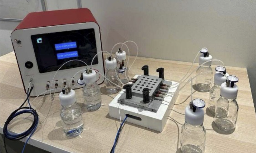
To date, prototypes of the MPS custom platform have been developed, characterised and provided to the project partners in the DeLIVERy project (Bielefeld, Tromso and Brussels). A representation of this platform can be seen in the figure to the right. These platforms aim to provide unprecedented biophysical parameter control to maintain physiologically relevant in-vitro models, modular imaging integration and multiplexed pharmaceutical compound testing. Ongoing platform testing and protocol refinement is underway at each of the partner sites (Bielefeld, Tromso and Brussels) for the complex 3D in-vitro LSEC models being developed within the project.
Microphysiological System (MPS) platform being developed on-site at Cherry Biotech